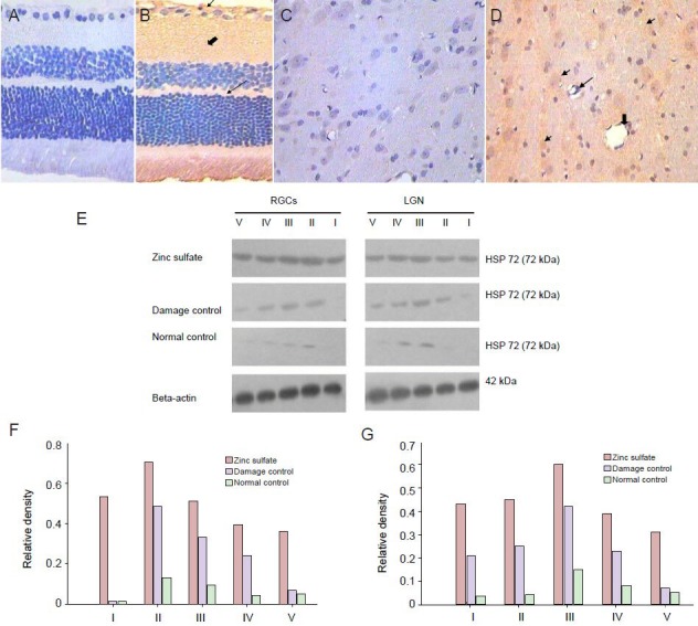
Figure 2.

Effect of laser treatment on the presence and expression of heat shock protein 72 (HSP72) in retinal ganglion cells (RGCs) and the lateral geniculate nucleus (LGN) of rats following different drug injections.
(A–D) Immunohistochemistry for HSP72. (A) HSP72 is not detected in the normal retina. (B) Compared with the normal control group, the pres-ence of HSP72 (brown staining) is less prominent at 7 days in the retinal ganglion cell layer (thin short arrow), nerve fiber layer (thick arrow) and outer plexiform layer (slender arrow) of the retina. (C) HSP72 is not detected in the normal LGN. (D) Seven days after laser treatment, LGN neurons are detected in tissues that label HSP72 (thin short arrow), vacuolated cells, nuclear condensation (thin arrow), and capillaries (thick arrow). West-ern immunoblot and quantitative analysis of HSP72 protein expression in the RGCs (E, F) and LGN (E, G) at different time points. At 3, 7, 14, and 28 days after laser treatment (II–V), HSP72 protein expression in the retina and LGN is significantly different at each time point (P < 0.05). I: Before treatment (damage control group).
