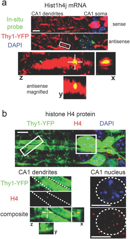
Figure 6. Dendritic localization of mRNA and protein encoded by the chromatin-associated gene Hist1h4j.

(a) In situ hybridization for Hist1h4j shows puncta within YFP-labeled dendrites with the antisense probe, but not with the sense probe (green = Hist1h4j probe, red = YFP, blue = DAPI). The bottom panels show magnified views of the area indicated by the white box. Views from all three planes show colocalization between Hist1h4j mRNA and the YFP-labeled dendrite. (b)Histone H4 IHC results in dendritically-localized puncta (green = Thy1-YFP, red = Histone H4, blue = DAPI). Magnified dendritic and somatic areas are indicated with white boxes. Dashed white lines in the magnified dendritic image outline a single dendrite. Views from all three planes show colocalization between Histone H4 protein and the dendrite. A dashed white circle in the magnified somatic image outlines a nucleus with the expected presence of Histone H4 protein. Scale bars = 10μm.
