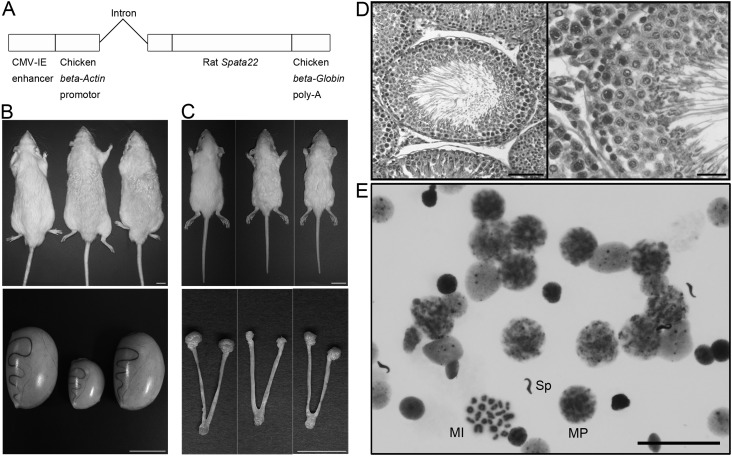
Fig. 4.
Recovery of spermatogenesis by transgenic rescue. A: Transgenic vector constructed by insertion of full-length rat Spata22 cDNA into pCAGGS. B and C: Gross examination of 7-month-old testis (B) and 21-day-old ovary (C) in mutants carrying the transgenes. Upper panels: dorsal views of animals. Lower panels: gross morphologies of gonads, oviducts, and uteri. In each panel, the genotypes of the animals and the organs are as follows: tm/+ (left), tm/tm (middle), and tm/tm Tg/+ (right). The homozygous mutant testis was small, and the homozygous mutant ovary could not be found despite the presence of oviducts and uteri. The testis and the ovary of the tm/tm Tg/+ rats appeared to develop normally, whereas the wavy coat phenotype of the tm/tm Tg/+ rats was similar to that of the tm/tm littermates in both sexes. Scale bar: 2 cm in upper panels, 1 cm in lower panels. D: Hematoxylin and eosin (HE)-stained section of testes from adult homozygous mutants that carry the transgenes, showing normal morphology of the seminiferous epithelium. Scale bar: 100 µm and 25 µm for the low- and high-magnification images, respectively. E: Giemsa-stained preparation of testicular cells, cell nuclei, or chromosomes from adult homozygous mutants carrying the transgenes, showing normal progression of meiosis. Spermatocyte nuclei at midpachytene and metaphase I stages and a spermatozoon head are indicated by MP, MI, and Sp, respectively. Scale bar: 50 µm.

