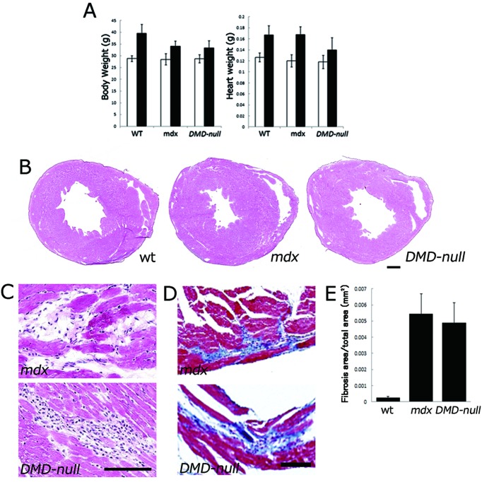
Fig. 4.
Phenotype analyses of cardiac muscle in mdx mice and DMD-null mice. A: Body weight (left panel) and heart weight (right panel) at 3 months (white bar) and 10–12 months (black bar) of age. B, C: Histological sections with hematoxylin-eosin staining. D: Histological sections with Masson’s trichrome staining. E: Graphic depiction of the ratio of connective tissue to normal myocardium in 12-month-old mdx, DMD-null mice and wt mice. The total area of blue-stained collagen was determined by digital image analysis. Note: No significant difference in necrotic area was observed between mdx and DMD-null mice (P=0.55).

