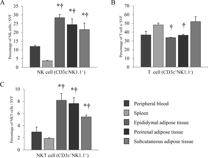
Fig. 2.
Lymphocytes fractions in adipose tissues, the spleen and peripheral blood. (A) NK cells (CD3ε−NK1.1+), (B) T cells (CD3ε+NK1.1−), and (C) NKT cells (CD3ε+NK1.1+) in SVFs from the spleen, peripheral blood and epididymal, perirenal and subcutaneous adipose tissues were evaluated by flow cytometry. SVFs derived from adipose tissues were obtained by removal of adipocytes from 20-week-old mice. Data represent means ± SEM (n=5).*P<0.05 vs. peripheral blood, †P<0.05 vs. spleen.

