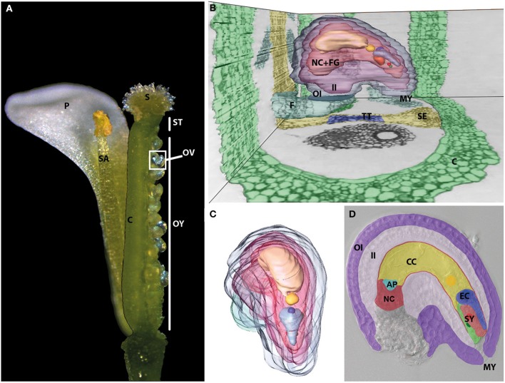
Figure 1.
The female gametophyte is deeply imbedded inside the female flower organs. (A) Dissected and reconstructed Arabidopsis flower. One of four petals (P) and one of six stamina (SA) are shown. They surround the pistil, which represents the female flower organ. It can be dissected into three parts. The upper part contains the papilla cells and forms the stigma (S), which is connected to the ovary (OY) by the style (ST). The ovary is formed by two fused carpels (C), which harbor two rows of ovules (OV). A side view (B) and front view (C) of a 3D-remodeled ovule reconstructed from toluidine blue stained single, successive ultra-thin sections of a dissected pistil. See Supplemental Movie 1 for whole series of sections. The ovule is connected to the septum (SE, yellow) containing the transmitting tract (TT, blue) by the funiculus (F, petrol) and surrounded by the carpel tissue (C) (green). A 3D-model of a dissected ovule shown from various angles is shown in Supplemental Movie 2. The mature female gametophyte cells (FG) and the nucellus tissue (NC) are surrounded by the outer (OI) and inner integuments (II) (OI, blue; II, purple). The vacuole and nucleus of the different female gametophyte cells showed highest contrast and are therefore shown individually. Near to the micropyle (MY), the two nuclei of the two synergid cells (SY) are shown in red and green. The egg cell, indicated by EC in (D), has a comparably large vacuole (light blue) and its nucleus (blue) is located at its chalazal pole. The center of the female gametophyte is filled by the vacuole (light yellow) of the central cell, indicated by CC in (D), and its homo-diploid nucleus (yellow). The three degenerating antipodal cells, indicated by AP in turquoise color in (D) at the chalazal pole are not highlighted. (D) DIC microscopic image of a mature female gametophyte surrounded by the maternal sporophytic tissues of the ovule. The cell types and tissues are artificially colored as shown in (B,C). At full maturity the nucellus cell (NC) layer surrounding the developing embryo sac is flattened between inner integument (II) and female gametophyte cells.

