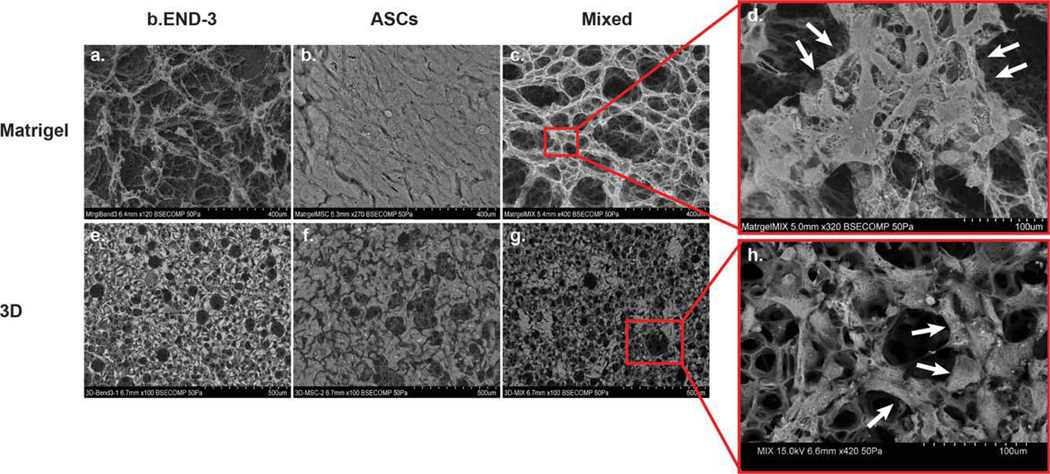
FIGURE 2.
SEM depicting organization and proliferation of ASCs, and b.END-3 cells when co-cultured or seeded alone on Matrigel and 3D polystyrene substrate (a–h). Scanning electron micrograph showing the multi-layered growth of ASCs (b and f) on both cultured surfaces. A construct made with ASCs, and b.END-3 cells, demonstrates the porous structure of the 3D scaffold (g). High-magnification image of a scaffold seeded with a ASCs, and b.END-3 cells, that demonstrate homogeneous and complete cellular distribution throughout the porous surfaces of the scaffold (h). The cells polarize around the pore (indicated by arrows) in a similar fashion as the one seen during the formation of branching, tubular structures (indicated by arrows developed in vitro in Matrigel (c and d). [Color figure can be viewed in the online issue, which is available at wileyonlinelibrary.com.]

