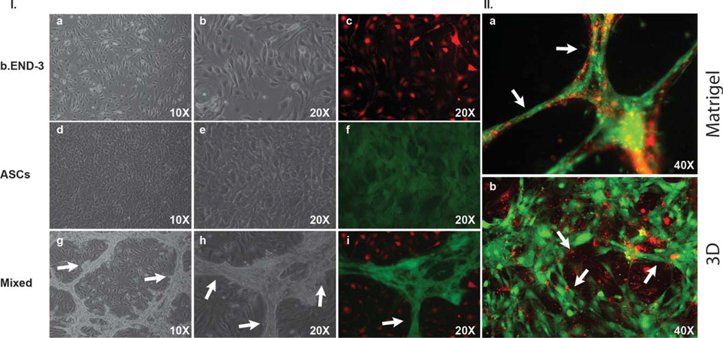
FIGURE 3.
Representative light and fluorescent microscopy images of ASCs, and b.END-3 cells when co-cultured or seeded alone on Matrigel or on 3D polystyrene substrate. Image of ASCs and b.END-3 on day 3 in the culture alone, demonstrating the absence of any well-formed tubular network l(a–f). Fluorescence microscopy images of GFP+ ASCs (green) co-seeded with PKH26-labeled b.END-3 cells (red) on Matrigel under normal conditions l(i). High magnification merged images show ASCs and b.END-3 cells working cooperatively in the formation of branching l(i). Completely lumenized tubular structrures develop in vitro in Matrigel 11(a). Note that the lumen is free of GFP+ and PKH26 signal 11(a). ASCs were seeded at a concentration of 1 × 10⁵ cells per well of 12-well plates and incubated for 72 h at 37°C in 5% C02. White arrows indicate tubule formation. Fluorescent microscopy of 3D scaffolds showing the 3D network of cell aggregates within the scaffold ll(b) arrows indicate lumen with ASCs contribute to in vitro formation of a 3D pre-vascular network within a porous scaffold. [Color figure can be viewed in the online issue, which is available at wileyonlinelibrary.com.]

