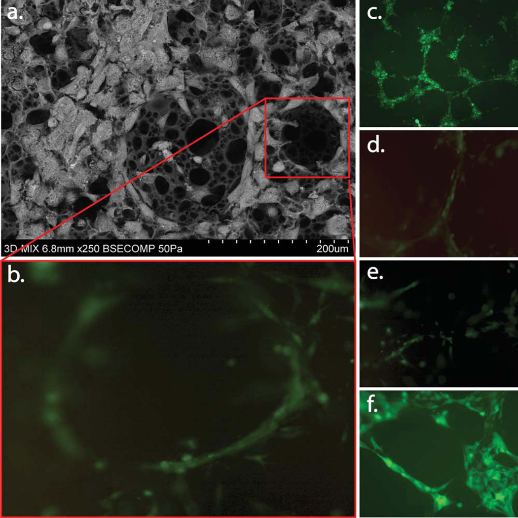
FIGURE 4.
Fluorescent microscopy of 3D scaffolds showing the 3D network of cell aggregates within the scaffold arrows indicate lumen with ASCs contribute to in vitro formation of a 3D pre-vascular network within a porous scaffold as seen in (c–f). SEM depicting organization and pro-iferation of ASCs, and b.END-3 cells when co-cultured on 3D polystyrene substrate (a). Physical interaction of ASCs with b.END-3 network formed in the 3D scaffold at 3 days post seeding. Confocal fluorescent images in (c–f) are cross-section projections at the surface (c) 60 µm, (d) 120 µm, (e) and 170 µm (f) within the polystyrene scaffolds. The penetration of the ASCs cells within the porous structures forming pre-capillary-like structures similar to the ones seen in matrigel control [Fig. 3(b,i)]. [Color figure can be viewed in the online issue, which is available at wileyonlinelibrary.com.]

