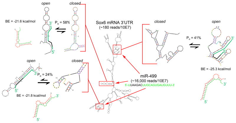
Figure 1. Schematic diagram of miR-499 binding to Sox6 mRNA.
An M-fold structure of Sox6 3′UTR is shown with miR-499 binding sites framed in red. These binding domains are enlarged as insets and shown in configurations less favorable (closed) and more favorable (open) for miR-499 seed sequence binding; probability of the more open structure is reported for each as Po. miR-499 is depicted on the open Sox6 configuration in green, with seed sequence in black; RNAhybrid duplex structure and minimum hybridization (binding) energy (BE) is given for each interaction. RNA mass/abundance values determined from publically available adult mouse heart RNA sequencing data are shown as reads/10 million reads.

