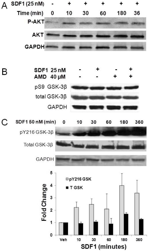
Figure 2. SDF1 stimulates AKT phosphorylation and increases overall GSK3β activity by Y216 phosphorylation in CSPCs.
A) CSPCs were treated with 25 nM SDF1 for increasing time periods in serum free media. CSPC homogenates were resolved by PAGE and phosphorylated and total AKT detected by Western analysis. Levels of total GSK3β and its S9 (B) and Y216 (C) phosphorylated forms were measured in homogenates after treating CSPCs with 25 nM SDF1 for 24 hours (Panel B) or 50 nM SDF1 for the specified time periods (Panel C) by Western analysis. Values are the mean ± SE, n=2-3.

