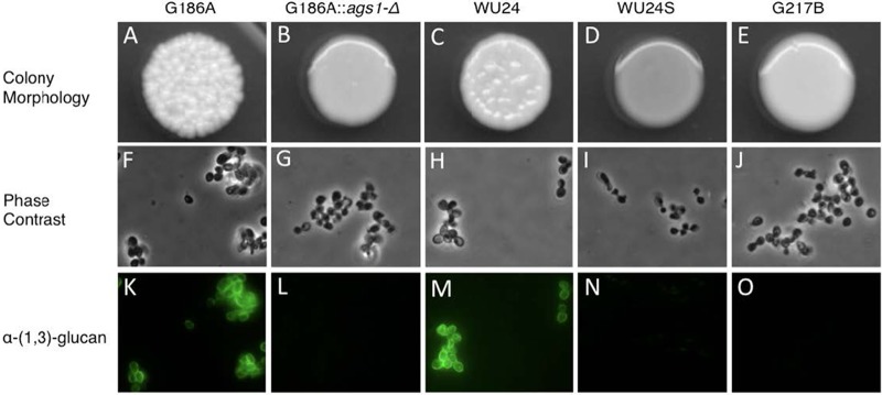
FIG 1 .

Colony morphology and α-(1,3)-glucan immunostaining. (A to E) Colony morphology of G186A (A) and WU24 (C), both chemotype II strains, which showed rough colony morphology, chemotype I strain G217B (E), which had smooth colony morphology, and also strains G186A ags1(Δ) (B) and spontaneous mutant WU24S (D). (F to J) Phase-contrast microscopy images of yeast broth cultures. (K to O) α-(1,3)-glucan immunostaining. The smooth colony morphology correlates with the absence of α-(1,3)-glucan in the yeast cell wall, as shown by immunofluorescence.
