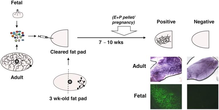
Fig. 1.
Schematic representation of the protocol for detecting fetal and adult MRUs. Cells from mammary glands are dissociated into a single-cell suspension and then transplanted into the cleared fat pad of a pubertal female mouse. Seven to ten weeks later, glands are removed and scored for the presence or absence of a large positive tree-structure. Photomicrographs show carmine-stained examples of positive and negative glands injected with adult cells (top) and examples of positive and negative glands injected with green fluorescent protein+ fetal cells (bottom). MRU detection can sometimes be increased by inducing pregnancy or implanting an estrogen and progesterone pellet (E + P pellet) 3 weeks prior to sacrifice

