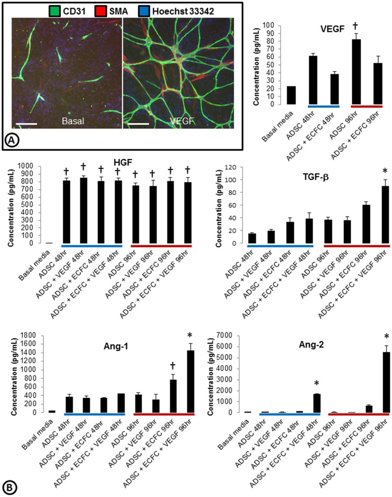
Figure 1. Characterization of growth factors present in the ADSC/ECFC co-culture system.
(A) Basal or VEGF driven co-cultures of ADSCs and ECFCs were stained for cords (CD31; green), smooth muscle actin (SMA; red), and nuclei (Hoechst 33342; blue). (B) Media was collected from ADSCs alone or ADSC/ECFC co-cultures in basal or VEGF driven conditions at 48 (blue bar) and 96 hrs (red bar). Examination of angiogenic growth factors present in the collected media was measured using Luminex. VEGF, HGF, TGF-β, Ang1, and Ang2 had detectable levels above basal media, while bFGF, EGF, and PDGF were not detected (data not shown). n = 3–4 per group with similar results found on two separate experiments. * = p<0.05 vs. all other treatment groups. † = p<0.05 vs. basal media. Scale bars are 250 µm.

