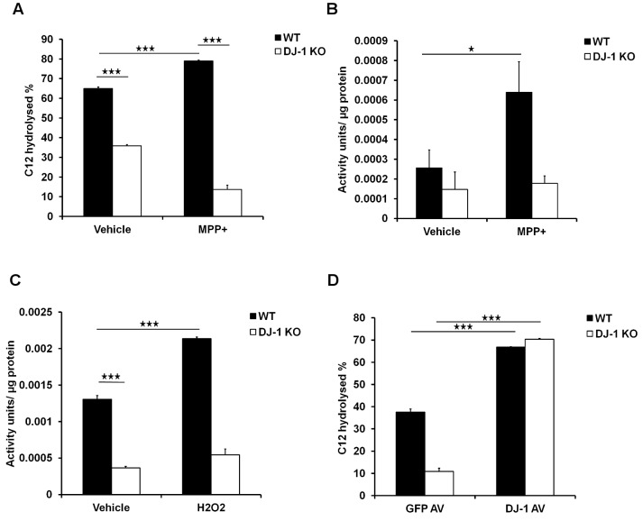
Figure 2. DJ-1 and oxidative stress modulate PON2 activity.
(A) Cultured WT and DJ-1 KO cortical neurons were treated with MPP+ (20 µM) for 12 hours and cells were washed and membrane was extracted. Crude membrane was exposed to the substrate C12 for 60 minutes and the percentage of remaining C12 was measured. (B) Cultured WT and DJ-1 KO cortical neurons were treated with MPP+ (20 µM) for 24 hours. Neurons were then exposed to DHC for 10 minutes and the amount of hydrolysis of DHC was assessed with measuring UV absorbance. One unit of PON2 activity is equal to 1 µmol DHC hydrolyzed/ml/min. (C) WT and DJ-1 KO MEFs were treated with hydrogen peroxide (100 µM) for 24 hours and PON2 activity was measured as described in B. (D) WT and DJ-1 KO MEFs were infected with adenovirus expressing DJ-1 or GFP alone as control. After 48 hours of expression, cells were lysed and exposed to C12 as the substrate for 60 minutes. Percentage of C12 remaining in activity buffer was measured. Statistical significance was assessed by Anova and post-hoc test Tukey on data obtained from three independent experiments (n = 3). * denotes p<0.05, ** denotes p<0.01, and *** denotes p<0.001.

