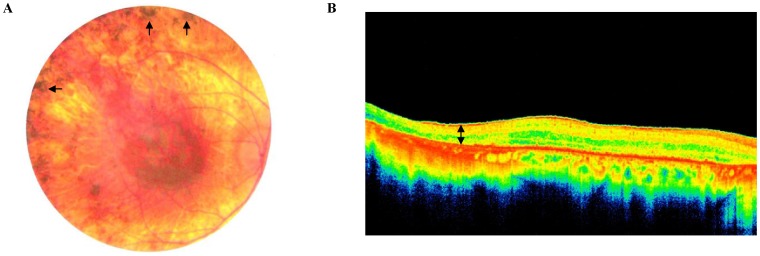
Figure 1. Clinical examination.
A. Posterior pole of the right eye of patient USHLB13-II.3 showing atrophy of the retina and choroid with pigment spicules (arrows) anterior to the arcades. B. Spectral domain optical coherence tomography of the right eye of the same patient showing significant thinning of the retina (space delineated by double arrow) compared to normal controls.

