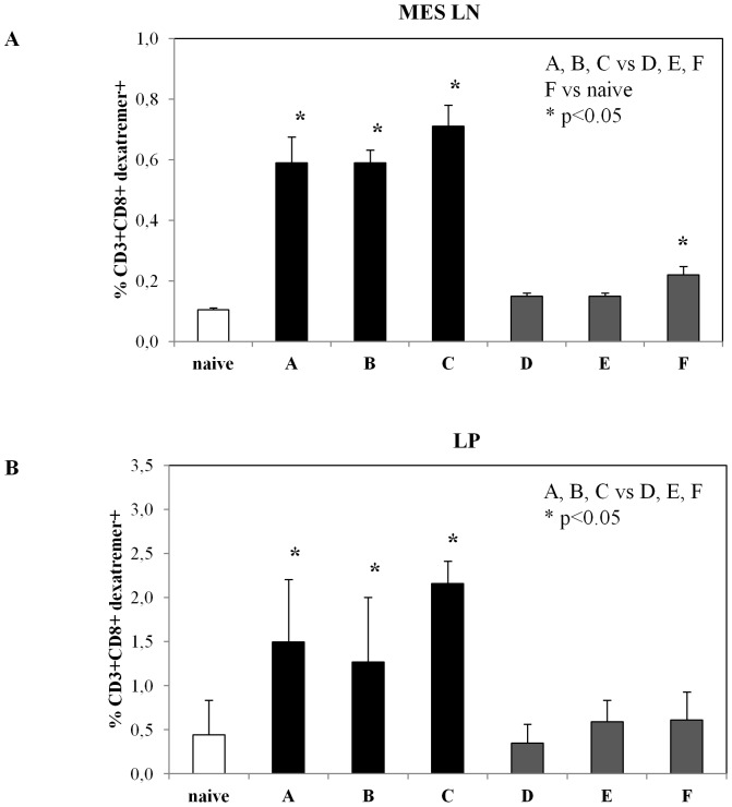
Figure 6. Persistence of mucosal antigen-specific CD8+ T cell response.
Six months after the last immunization mice from all groups (vaccination regimens are described in Table 1) were sacrificed. (A) Mesenteric lymph node-derived lymphocytes were stained with H-2Kb-SIINKFEL dextramers, anti-mouse CD3 and anti-mouse CD8. Results are expressed as percentage of CD3+CD8+ dextramers+ cells presented as group means ± standard deviations. The asterisks indicate statistically significant differences (p<0.05) between indicated groups. (B) Lymphocytes derived from large intestine lamina propria (LP) of mice from indicated group were stained with fluorescent H-2Kb-SIINKFEL dextramers, anti-mouse CD3 and anti-mouse CD8. The analysis was performed on gated CD3+CD8+ cells from immunized or naïve mice. Results are expressed as percentage of CD3+CD8+ dextramers+ cells presented as group means ± standard deviations. The asterisks indicate statistically significant differences (p<0.05) between indicated groups.

