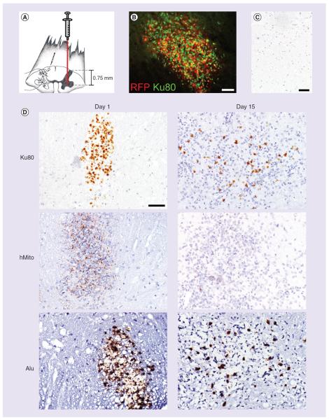
Figure 5. Detection of human glial-restricted precursors injected into the mouse spinal cord.
(A) RFP prelabeled human glial-restricted precursors (GRP) are injected into the mouse spinal cord, targeting the ventral horn. (B) At 1-day postinjection, RFP+ human GRPs are visualized in the injection site of the spinal cord. These cells co-express the human-specific biomarker Ku80. (C) Using anti-human Ku80, hMito and Alu immunohistochemistry, human GRP cells can be tracked in the spinal tissue from 1 day to 15 days post-transplantation. (D) Vehicle-injected animals do not show any biomarker immunoreactivity in the spinal tissue. While Ku80 and Alu immunoreactive cells are still found at 15 days post-transplantation, the signal for hMito is very weak. Scale bar represents 50 μm.
hMito: Human mitochondria; RFP: Red fluorescent protein.

