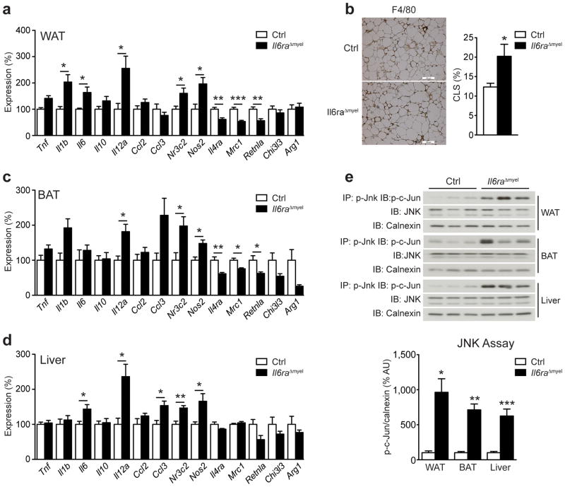
Figure 2. HFD Il6raΔmyel mice have increased systemic inflammation.
(a) qRT-PCR analyses of white adipose tissue (WAT) from HFD control (Ctrl) and Il6raΔmyel mice (n=8–9 per genotype; *p≤0.05; **p≤0.01; ***p≤0.001; Data are expressed as % of Ctrl). (b) Representative immunohistochemical staining of F4/80-positive cells in WAT from HFD Ctrl and Il6raΔmyel mice and quantification of F4/80-positive crown-like structures (CLS) in WAT from HFD Ctrl and Il6raΔmyel mice (n=7 per genotype;; *p≤0.05; Data are expressed as % CLS of adipocytes; scale bars represent 100μm). (c) qRT-PCR analyses of brown adipose tissue (BAT) from HFD Ctrl and Il6raΔmyel mice (n=8–9 per genotype; *p≤0.05; **p≤0.01; Data are expressed as % of Ctrl). (d) qRT-PCR analyses of liver from HFD Ctrl and Il6raΔmyel mice (n=8–9 per genotype; *p≤0.05; **p≤0.01; Data are expressed as % of Ctrl). (e) C-Jun N-terminal kinase (JNK) activity in WAT, BAT and liver of HFD Ctrl and Il6raΔmyel mice was measured by performing immunoprecipitation (IP) of p-JNK and after subsequent in vitro phosphorylation assay, recombinant c-Jun was detected by immunoblot (IB). Total JNK and calnexin loading was used as input control; Representative immunoblots are shown (upper panel); Quantification of JNK activity in WAT, BAT and liver of HFD Ctrl and Il6raΔmyel mice (lower panel; n=5–9 per genotype; *p≤0.05; **p≤0.01; ***p≤0.001; Data are expressed as % of Ctrl). (Values are expressed as mean ± sem;).

