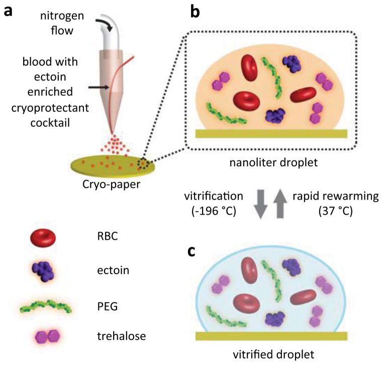
Figure 1. Droplet-based vitrification of red blood cells.
a, The essentials of the experimental setup for the droplet formation. The system consists of nitrogen gas flow and a droplet deposition system controlled by a syringe pump. The droplets are generated from the co-flow stream of cryoprotective agent (CPA)-loaded red blood cell (RBC) mixture as the nitrogen gas flows through a droplet ejector, which transforms the bulk of the sample into nanoliter droplets. The CPA consists of a cocktail solution of ectoine, trehalose, and polyethylene glycol (PEG). The droplets were ejected on a cryo-paper (polyethylene collection film). b, The magnified view of a single droplet on the cryo-paper including RBCs, ectoine, trehalose and PEG. c, The view of the vitrified droplet in (b). The vitrification is achieved by submersing the cryo-paper into liquid nitrogen. The vitrified droplet transforms to (b) after rapid thawing of the cells on a cryo-paper in phosphate-buffer saline at 37 °C.

