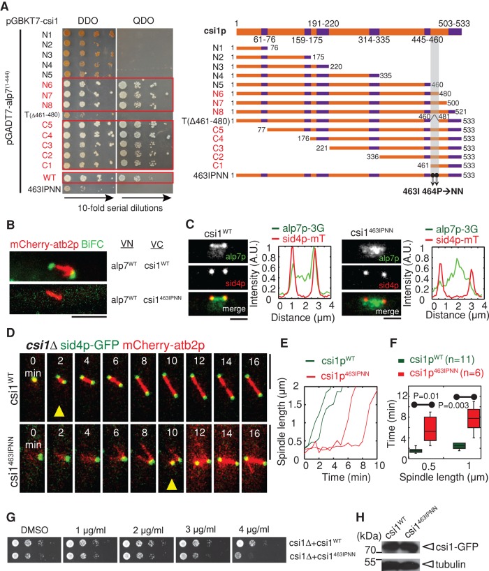
FIGURE 4:
Csi1p carboxyl terminus is responsible for interacting with alp7p. (A) Yeast two-hybrid assays for mapping the minimal alp7p interaction domain in csi1p. A series of csi1p deletion truncation mutants, as indicated in the schematic diagram (coiled-coil domains indicated in purple), was used to test their interaction with alp71-444, revealing that domain 461–480 at the csi1p carboxyl terminus is important for interacting with alp71-444. Further, the two residues Ile-463 and Pro-464 within the minimal domain in csi1p are key residues responsible for the interaction with alp7p. (B) BiFC assays. Maximum projection images of cells expressing alp7pWT-VN and csi1pWT-VC or csi1p463IPNN-VC from the nmt1 promoter. Cells were cultured in EMM (Edinburgh minimal medium) without thiamine for 14 h before imaging. Note that only the cell expressing wild-type csi1p gave BiFC signals. Scale bars, 5 μm. (C) Maximum projection images of csi1∆ cells expressing alp7p-3GFP, sid4p-mTomato, and either wild-type csi1p (indicated as csi1WT) or mutant csi1p463IPNN (indicated as csi1463IPNN) from a csi1p promoter at the leu1-32 locus. Fluorescence intensity measurements were carried out to analyze alp7p signal profiles along the spindles. Alp7p no longer concentrated at the SPBs in the csi1p463IPNN cell. Scale bars, 2 μm. (D) Maximum projection live-cell images of csi1WT and csi1463IPNN cells expressing sid4p-GFP and mCherry-atb2p. Yellow triangles mark bipolar spindles ∼1 μm in length. Note that the csi1p463IPNN cell displayed transient monopolar spindle formation. Scale bars, 5 μm. (E) Representative plots of spindle length against time for csi1WT and csi1463IPNN cells. (F) Box plots of time for assembly of bipolar spindles measuring 0.5 and 1 μm in length in csi1WT and csi1463IPNN cells. Student's t test was used to calculate p values. Cell numbers analyzed are indicated. (G) MBC sensitivity assays for csi1WT and csi1463IPNN cells. The cells were grown at 30°C for 4 d. Similar to csi1∆, csi1463IPNN cells were sensitive to MBC. (H) Western blot analysis of cells expressing csi1WT-GFP and csi1463IPNN-GFP.

