
FIGURE 4:
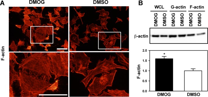
PHD3 inhibitor DMOG increases β-actin polymerization. HeLa cells were treated with DMSO or DMOG (500 μM) for 72 h. (A) Cells were fixed, permeabilized, stained with Alexa Fluor 555–conjugated phalloidin, and imaged by fluorescence microscopy. The boxed areas are enlarged and shown below. Representative images from at least three independent experiments. Scale bar, 100 μm. (B) Actin sedimentation assays were performed, followed by immunoblot assays with anti–β-actin antibody. F-actin bands were quantified by densitometry and normalized to DMSO (mean ± SEM, n = 3). *p < 0.05 vs. DMSO.