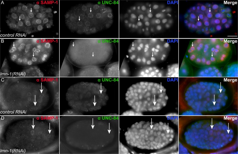
FIGURE 7:
SAMP-1 localizes independently of LMN-1. (A–D) Embryos were stained for SAMP-1 and UNC-84 localization. Lateral views, with anterior left and dorsal up. For each row, SAMP-1 immunostaining is shown in white in the left column and in red on the right when all channels are merged. UNC-84 is shown in white in the second column from the left and in green when merged. DAPI staining of nuclei is shown in white in the third column and in blue when merged. (A) An early embryo fed bacteria containing the empty L4440 vector as control. (B) An early embryo fed lmn-1(RNAi). (C) A later, pre–comma-stage embryo fed bacteria containing the empty L4440 vector as control. (D) A later, pre–comma-stage embryo fed lmn-1(RNAi). Arrows highlight specific nuclei to provide reference points in all four columns. Scale bar, 10 μm.

