
Figure 5.
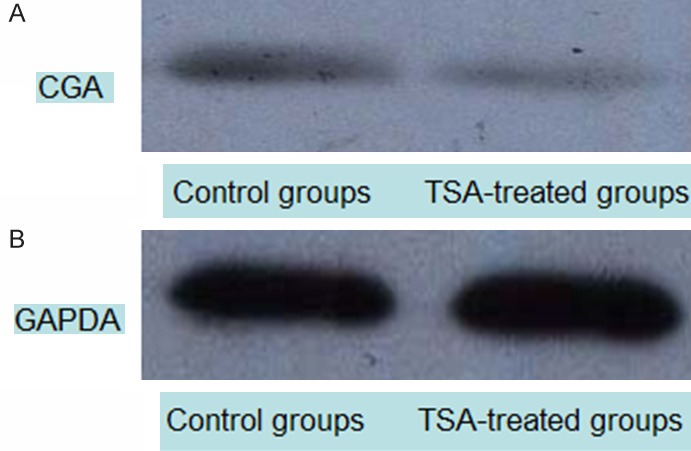
CGA protein levels detected by western blot. Cell lysates were analyzed by Western blot using anti-GAPDH and Anti-CGA antibodies. A: CGA (56 KDa); B: GAPDH (loading control, 37 KDa). CGA protein expression was reduced in the TSA group at 48 h compared with controls.