
Figure 6.
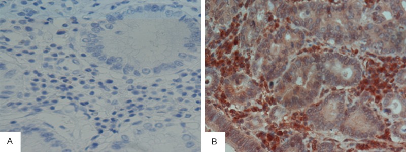
CGA detection by immunohistochemistryin tissue specimens from gastric adenocarcinoma patients. Expression of CGA was mainly localized tothe cytoplasm. Based on a method combining the percentage of positive cells and signal intensity, 74% were positive for CGA expression in gastric adenocarcinoma tissue samples. In normal tissue specimens adjacent to carcinoma, only 24% were deemed positive. This indicates a significant difference in CGA expression between gastric adenocarcinoma than normal tissues (A: Negative, B: Positive; ×400).