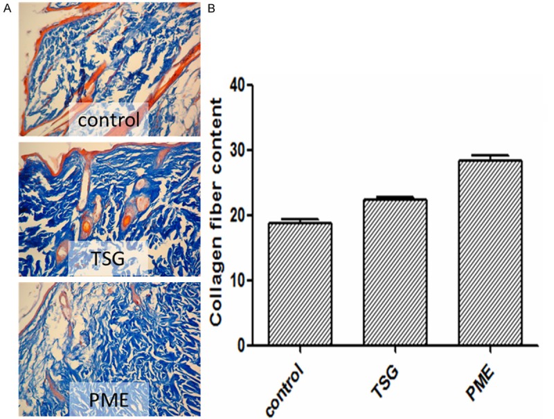
Figure 2.

A. TSG and PME increased the thickness of dermal layer (×200). Collagen fiber was shown by blue color in three pictures which proved that TSG and PME groups had larger distrubution and more orderly than the control group. B. The result of image scan showed more cleared that the contant of collagen fiber in TSG and PME groups were significantly more than control group (p<0.01, vs. Control group).
