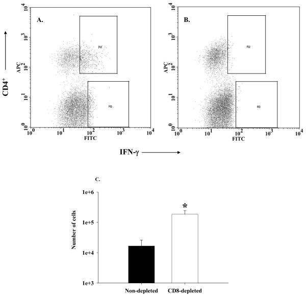
Figure 6. CD4+ IFN-γ+ T cells are readily detected in the lung tissue of experimental mice.
Lung cells were isolated from experimental mice sacrificed on the seventh day after immune reconstitution, stimulated with PMA and ionomycin, and stained with fluorescent MAbs against the following surface markers: CD4 (APC) and CD8 (PerCP) as well as intracellular IFN-γ (FITC). Results of pooled cells from CD8-depleted IRD mice are shown in Panel A. The same cells pre-treated with 10 µg of unlabeled anti-IFN-γ shows nearly complete blocking of IFN staining. The percentage of CD4+ IFN-γ+ cells [cells in the gate R2] from a similar plot for each sample were used to calculate total number of IFN-γ -producing CD4+ T cells in that sample, and the results are shown in Panel C. Bars represent arithmetic means of three samples and error bars represent 1 SEM *p<0.05..

