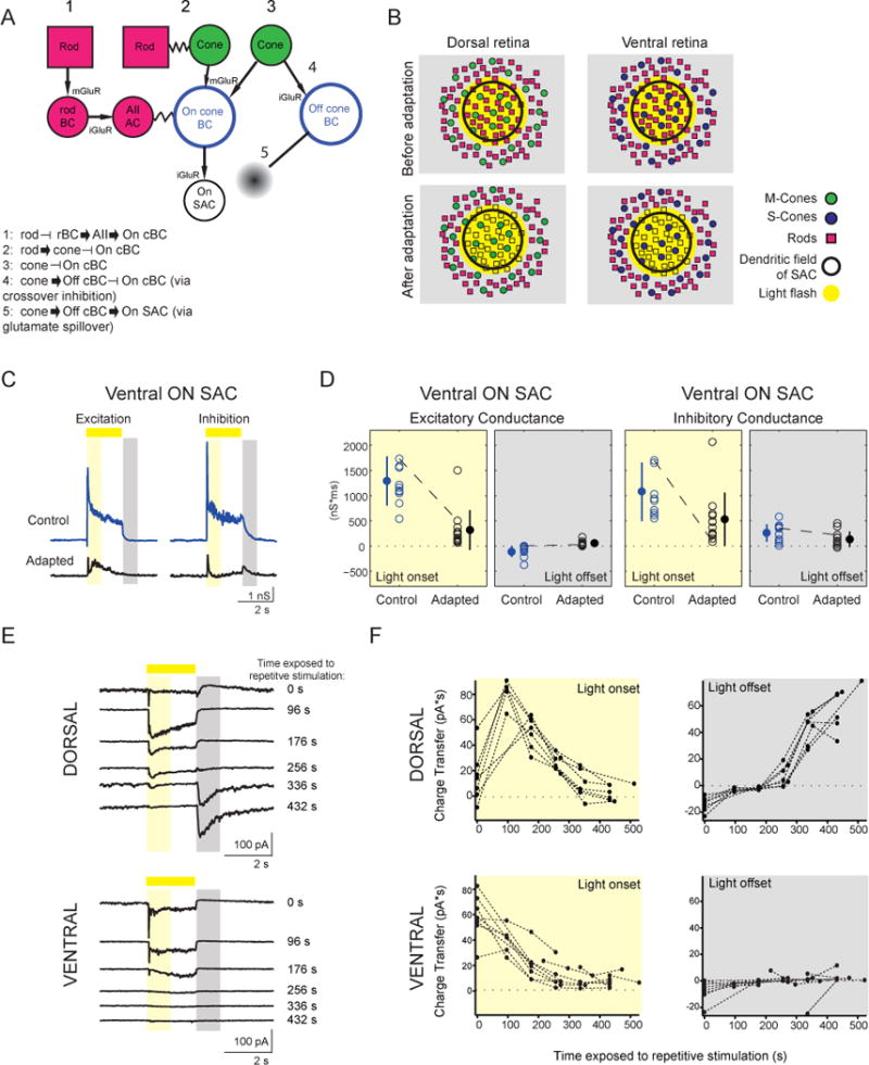
Figure 4. Gain of Off Response Requires Cone Activation.

(A) Schematic of potential pathways that could influence signaling in On-SACs.
(B) Schematic of experimental results. Light stimulation with an OLED activated both rods and cones in the dorsal retina and only rods in ventral retina. Following repetitive stimulation, rods are no longer responsive to light spots.
(C) Excitatory and inhibitory conductances from a ventral On-SAC. Conventions are as in Figure 2C.
(D) The integrated excitatory and inhibitory conductances during light onset and light offset for ventral On-SACs before and after adaptation. Conventions are as in Figure 2E.
(E) Voltage clamp recordings of excitatory currents from dorsal (top) and ventral (bottom) On-SACs (holding potential = −72 mV) in response to a 2-s light spot (225 μm diameter). Spots were presented in between exposing the cells to repetitive stimulation, with the time exposed to repetitive stimulation indicated on the right. Traces are averages of five sweeps. The time periods for calculating the charge transfer are indicated by the yellow rectangle for light onset (50–850 ms after light onset) and by the grey rectangle for light offset (100–900 ms after light offset).
F, Charge transfer (averaged over five sweeps) of the excitatory current during light onset and light offset for dorsal (top) and ventral (bottom) On-SACs as a function of the amount of time cells were exposed to gratings.
See also Figure S3.
