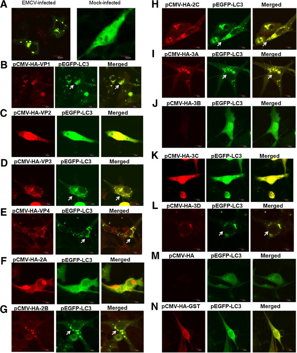
Figure 1.

Immunofluorescence puncta in the cytoplasm of BHK-21 cells that were co-transfected with each HA-tagged EMCV protein-expressing plasmid and pEGFP-LC3. (A) Confocal immunofluorescence analysis was performed on BHK-21 cells transfected with the pEGFP-LC3 plasmid at 18 h after transfection, and the cells were then infected with the EMCV BJC3 strain for 12 h (MOI = 0.005). A GFP-LC3 signal (green) and EMCV VP1 protein staining (red) are shown. (B-N) The BHK 21 cells were co-transfected with each HA-tagged EMCV protein-expressing plasmid or GST protein-expressing plasmid and pEGFP-LC3. At 48 h after transfection, the cells were fixed and processed by immunostaining with a mouse monoclonal antibody against HA and goat anti-mouse secondary antibodies conjugated to TRITC. The HA (red) and GFP-LC3 (green) proteins were examined by immunoconfocal microscopy. The white arrows show the positive puncta of LC3 proteins or the colocalization of the LC3 proteins and EMCV proteins. The scale bars in panels (A-N) represent 10 μm.
