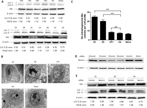
Figure 2.

Nonstructural proteins 2C and 3D increased autophagic activity and the formation of autophagosomes. (A) Western blotting analyses of LC3, p62 and β-actin in BHK-21 cells that were transfected individually with an HA-tagged EMCV protein-expressing plasmid. HA, the BHK-21 cells transfected with pCMV-HA; Mock, the untransfected BHK-21 cells. The band intensities of LC3-II, p62 and β-actin were quantified, and the relative ratios of LC3-II/β-actin and p62/β-actin are shown in the lower blots. (B) The BHK-21 cells were transfected with pCMV-HA-2C, pCMV-HA-3D, pCMV-HA-VP1 or pCMV-HA and mock (untransfected BHK-21 cells) for 48 h, and they were fixed, processed, and imaged by transmission electron microscopy (TEM). The morphologically characteristic double-membrane vesicles are indicated by black arrows in the relevant areas. Magnification, 10,000×; scale bars, 2 μm. (C) A quantification of the number of autophagosome-like vesicles per cell profile in BHK-21 cells that were transfected with 2C-, 3D-expressing plasmid, pCMV-HA-VP1 or pCMV-HA and mock (untransfected BHK-21 cells) Data are the means ± SD (error bars) for 8 cells per experimental condition from three independent experiments. ***p < 0.001, compared with the control cells. (D) BHK-21 cells were transfected with 10, 20, 30 or 40 pmol Beclin1-siRNA or 40 pmol control-siRNA, and then Beclin1 protein was detected by western blotting at 48 h post-transfection. Mock, the untransfected BHK-21 cells. (E) BHK-21 cells transfected with 40 pmol Beclin1-siRNA or control-siRNA. After 48 h, the knockdown cells were reseeded, transfected with pCMV-HA-2C, pCMV-HA-3D or pCMV-HA for an additional 48 h and processed for Western blotting analysis. The band intensities of LC3-II, p62 and β-actin were quantified, and the relative ratios of LC3-II/β-actin and p62/β-actin are shown in the lower blots.
