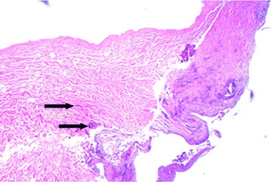
Fig. 6.

Hematoxylin and eosin staining of biopsy from patient #1 following 16 weeks in situ placement of acellular dermal matrix. Note the intact ultrastructure and also evidence of cellular in-growth as apparent fibroblasts (arrows) at ×10 magnification
