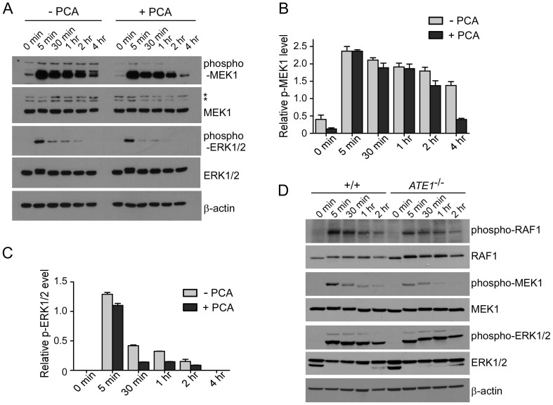
Figure 4. PCA impairs the downstream activations of GPCR signaling.
(A) Activation profiles of MAPK signaling proteins in HeLa cells in the presence or absence of 100 μM PCA. Cells were incubated with serum-free media for 12 hr, followed by serum-containing media. Samples were collected at the indicated time points and analyzed by SDS-PAGE/IB. (B & C) Quantification of phospho-MEK1 (B) and phospho-ERK1/2 (C) normalized by total MEK1 and ERK1/2, respectively, in the presence/absence of PCA. Multiple film images were quantified by densitometry and data represent mean ± SD (n = 3). (D) As in (A) except that the experiment was performed using wild-type (+/+) and ATE1-/- MEFs. Cropped gels/blots are used in Western blot data.

