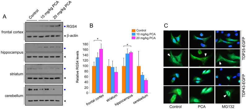
Figure 6. PCA delays RGS4 degradation in the frontal cortex and hippocampus while facilitates aggregation formation of Arg208-TDP25.
(A) Mouse brains after control and PCA injection were dissected into the frontal cortex, striatum, hippocampus, and cerebellum. Then samples were extracted and used for RGS4 immunoblotting. Cropped gels/blots are used here. (B) Quantification of RGS4 levels in brains normalized by β-actin, which indicate significantly increased levels of RGS4 in the frontal cortex and hippocampus. Mean ± SD (n = 4/group). (C) TDP43-EGFP and Arg208-TDP25-EGFP was transfected into HeLa cells which were treated with MG132 (10 μM) or PCA (100 μM) for 4 hr after 36 hr post-transfection.

