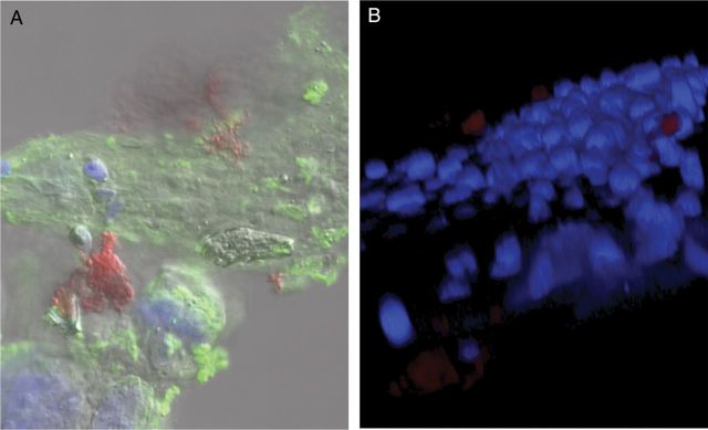
Figure 6.

Confocal analysis of MGAS315 biofilm colonization of nasal-associated lymphoid tissue (NALT). Eight-week old female BALB/cByJ mice were infected intranasally with biofilm MGAS315 that was formed for 48 hours on paraformaldehyde-fixed squamous cell carcinoma (SCC) 13 cells. At 72 hours after inoculation, mice were euthanized, and NALT was removed and fixed in 4% neutral buffered formalin. A, NALT was stained with 488-conjugated wheat germ agglutinin (green), nuclei stained with 4′,6-diamidino-2-phenylindole (DAPI), and group A streptococcus (GAS) stained with anti-GAS antibody (red). B, Representative 3-dimensional reconstruction of the specimen, using confocal images stained with DAPI (blue) and anti-GAS antibody (red).
