Figure 1.

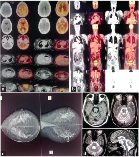
(a and b) PET-CT Brain and whole body showing absence of obvious FDG –avid lesion, (c) Mammography – Normal, D; T1W axial, T2W axial and sagittal section brain showing cerebellar atrophy

(a and b) PET-CT Brain and whole body showing absence of obvious FDG –avid lesion, (c) Mammography – Normal, D; T1W axial, T2W axial and sagittal section brain showing cerebellar atrophy