Figure 1.
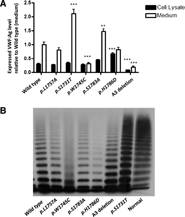
Expression of recombinant mouse VWF:CB variants. (A) Transient transfection of murine VWF cDNA was performed in HEK293T cells and expressed in serum-free medium. Total VWF:Ag level in medium and lysates per 10-cm dish was measured via VWF:Ag ELISA and results were normalized to WT-VWF expressed in medium equal to 1 (n = 10). *P < .05, **P < .01, ***P < .001. (B) Multimeric analysis for r-mVWF. All of the mutant mVWFs had a normal complement of HMWMs, except p.W1745C, which showed a smeary multimer pattern. The A3 deletion mutant showed a triplet band shift corresponding to loss of the A3 domain.

