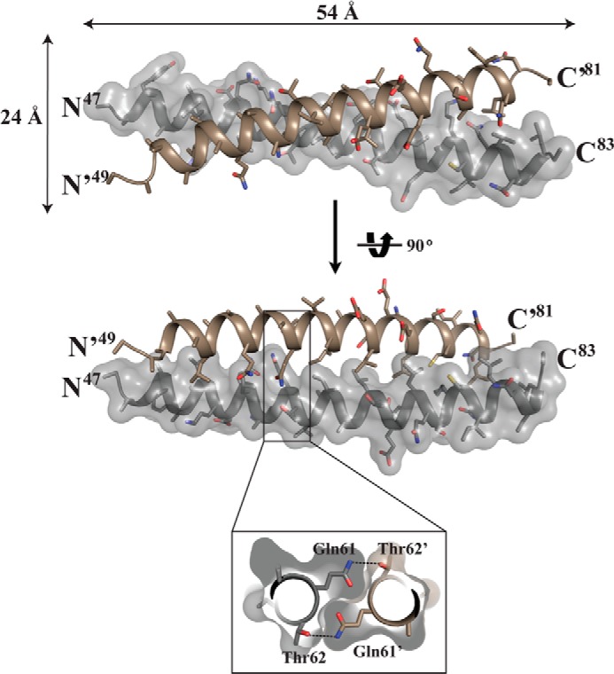
FIGURE 3.

Structure of the PKG II LZ domain. The structure of the PKG II LZ domain showing the overall dimensions in angstroms (Å), with stick representation of the side chains from each monomer. Only one of the chains is shown with surface. Inset, cutaway view looking down the 2-fold axis. This view shows the symmetrical hydrogen bond formed by Gln-61 of one chain and Thr-62 of the other.
