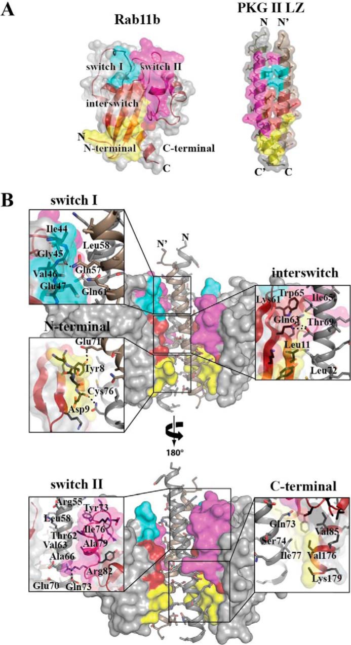
FIGURE 6.

Interface of the PKG II LZ-Rab11b complex. A, color-coded representation of the regions on Rab11b and PKG II that form the interaction interface. The coloration corresponds to the Rab11b surfaces that interact between the two proteins: switch I (teal), switch II (magenta), interswitch (red), and N terminus (yellow). B, PKG II LZ-Rab11b interactions with zoom-in views. Rab11b provides the largest interface at the interswitch/N-terminal region, contributing van der Waals contacts from residues Asp-9 to Phe-12, Phe-48, Lys-58, Lys-61, Trp-65, and Val-85. PKG II LZ provides residues Thr-62, Ile-65, Ala-66, Leu-72, Gln-73, Cys-76, Ile-77, and Lys-81 of PKG II LZ and Gln-61′, Ala-64′, Glu-67′, and Leu-68′ of PKG II LZ′ to this interface. Dashed lines in the insets represent hydrogen bonds and salt bridge interactions.
