FIGURE 7.
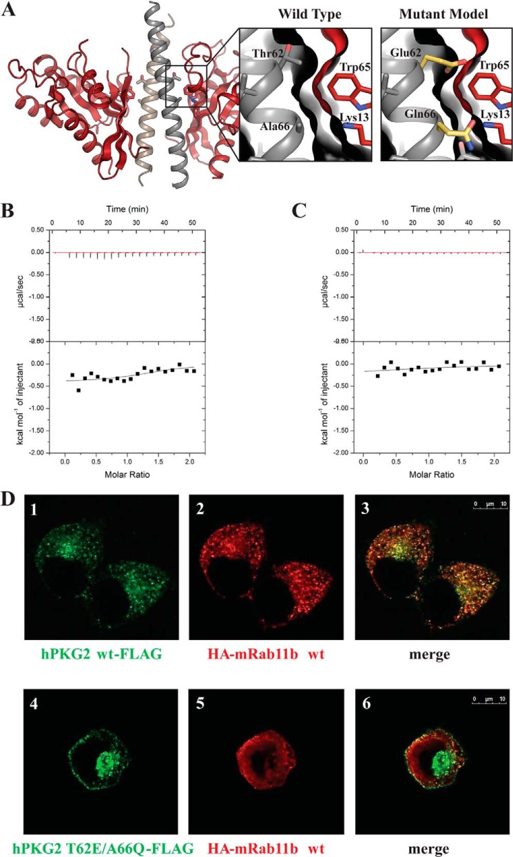
Representation of mutations made to Rab11b, mutant ITC measurements, and subcellular localizations of PKG II and Rab11b in HeLa cells. A, schematic representation of the PKGII LZ-Rab11b complex with enlarged panels showing the interface seen in the crystal structure (left) and in a modeled structure with PKG II T62E/A66Q mutations (right). B and C, ITC was performed as described under “Experimental Procedures.” No exothermal response was seen when titrating Rab11b into PKG II LZ with a T62E mutation (B) or A66Q mutation (C). D, HeLa cells expressing PKG II-FLAG (panels 1–3) or PKG II T62E/A66Q-FLAG (panels 4–6) together with HA-Rab11b were analyzed by confocal microscopy, as described under “Experimental Procedures.” PKG II WT-FLAG (panel 1, green) and HA-Rab11b (panel 2, red) co-localized in the recycling compartment (panel 3, areas of colocalization are yellow), whereas PKG II T62E/A66Q-FLAG did not co-localize with HA-Rab11b (panels 4–6).

