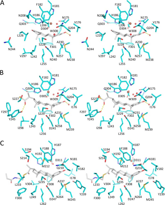
FIGURE 7.
Stereo substrate-binding pockets of KshAMtb·ADD (A), KshA1·1,4-BNC-CoA (B), and KshA5·1,4-BNC-CoA (C). The carbon atoms of the enzyme and steroid are shown in cyan and gray, respectively. Nitrogen, oxygen, and sulfur are dark blue, red and yellow, respectively. The mononuclear iron is a brown sphere and waters are red spheres. H-bonds are shown as yellow dashed lines. Figure was generated using PyMOL (50).

