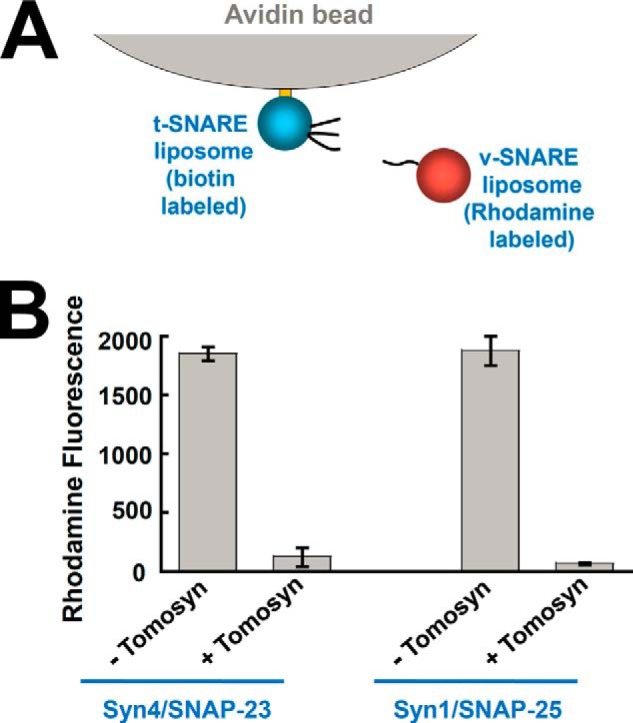
FIGURE 5.

Tomosyn blocks SNARE-mediated liposome docking. A, diagram of the SNARE-liposome docking assay. B, biotin-labeled t-SNARE liposomes were anchored to avidin beads and were used to pull down rhodamine-labeled v-SNARE liposomes. The binding reactions were performed in the absence or presence of 5 μm tomosyn. The binding reaction containing 20 μm VAMP2 CD was used as a negative control to obtain the background fluorescent signal. The background fluorescence was subtracted from other binding reactions to reflect specific SNARE-dependent liposome docking. The data are presented as rhodamine fluorescence intensity. Error bars indicate S.D.
