FIGURE 6.
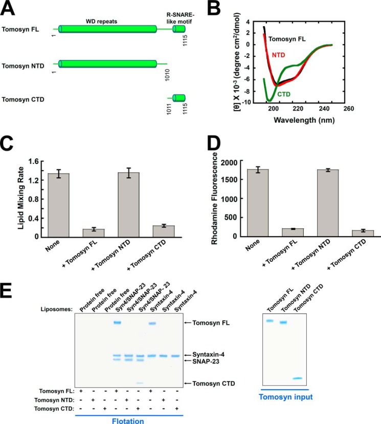
Tomosyn uses its CTD to arrest membrane fusion whereas its NTD is necessary for binding to syntaxin monomer. A, diagrams of tomosyn FL, as well as the NTD and CTD of tomosyn. B, CD spectra of tomosyn and tomosyn fragments. C, initial lipid mixing rates of the indicated SNARE-mediated fusion reactions in the absence or presence of 5 μm tomosyn FL or tomosyn fragments. Each fusion reaction contained 5 μm t-SNAREs and 1.5 μm v-SNARE. Data are presented as percentage of fluorescence change per 10 min. Error bars indicate S.D. D, tomosyn NTD does not block SNARE-mediated liposome docking. The binding reactions were performed in the absence or presence of 5 μm tomosyn FL or tomosyn fragments. Error bars indicate S.D. E, left: Coomassie Blue-stained SDS-PAGE gel showing the binding of tomosyn FL and tomosyn fragments to the t-SNARE liposomes reconstituted with syntaxin-4/SNAP-23 or syntaxin-4 monomer. The liposomes were incubated with tomosyn FL or tomosyn fragments at 4 °C for 1 h, followed by flotation on a Nycodenz gradient. Each binding reaction contained 5 μm SNAREs. Soluble factors were added to a final concentration of 5 μm. Right, Coomassie Blue-stained SDS-PAGE gels showing recombinant tomosyn FL and tomosyn fragments.

