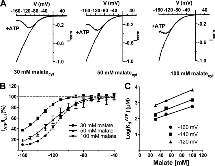
FIGURE 2.
Cytosolic ATP compete with cytosolic malate to inhibit AtALMT9-mediated currents. A, normalized current-voltage curves obtained with a voltage ramp protocol (from +60 to −160 mV in 1. 5 s) in cytosolic-side buffers containing various concentrations of malate (30, 50, and 100 mm malatecyt) with constant vacuolar side conditions (100 mm malatevac) in the presence (+ATP) or absence of 1 mm ATPfree. B, fraction of unblocked current in the presence of 1 mm ATPfree (IATP/Ictrl; mean ± S.E., n = 4–7) plotted against the applied voltage in different cytosolic malate concentrations. Data were fitted with Equation 3 (“Experimental Procedures”; Table 1; solid line). C, logarithmic plot of the dissociation constant for ATPfree (KdATP) at different applied membrane potentials (−120, −140, −160 mV) shown as a function of different cytosolic malate concentrations (30, 50, and 100 mm malatecyt). Data were fitted with a straight line with no theoretical significance to extrapolate the value of the dissociation constant at malate concentrations in the physiological range. Error bars denote S.E.

