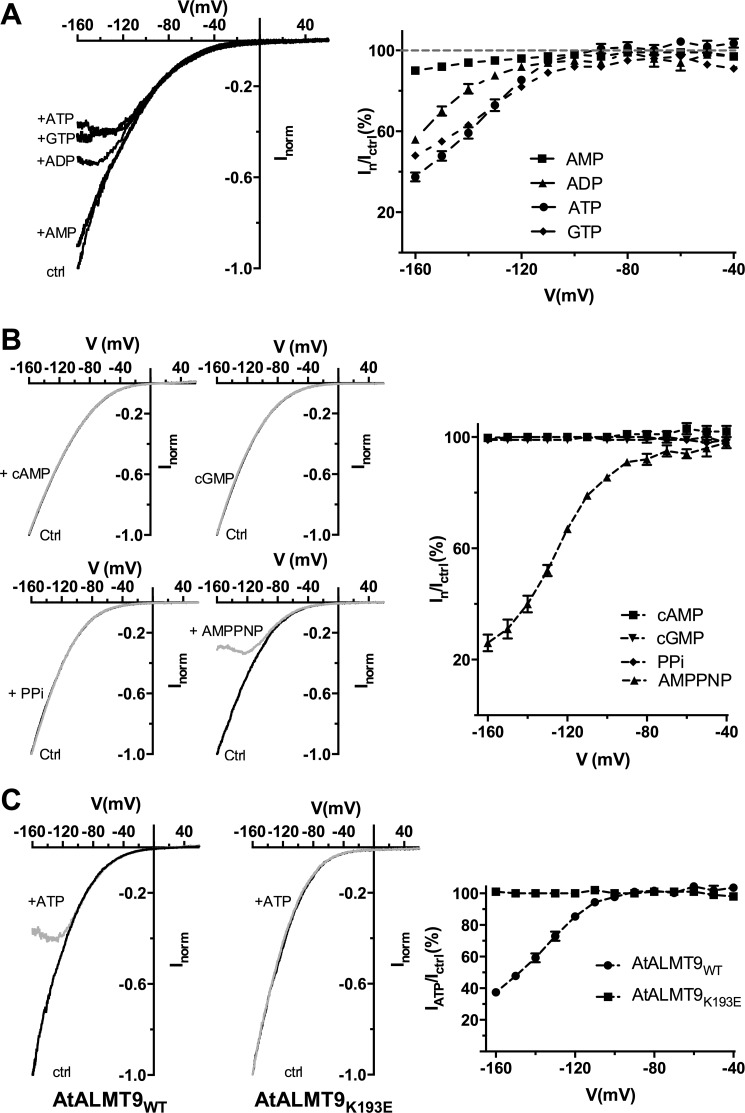
FIGURE 5.
Effect of the non-hydrolysable nucleotide AMPPNP and different cytosolic nucleotides on AtALMT9-mediated malate currents. A, left panel: representative currents elicited by a voltage ramp (from +60 to −160 mV in 1.5 s, holding potential +60 mV) in 100 mm malatecyt, 100 mm malatevac (ctrl), and in 100 mm malatecyt, 100 mm malatevac + 1 mm of different nucleotides in the cytosolic-side buffer: ATP, ADP, AMP, GTP. Right panel: mean current ratios between currents in the presence of 1 mm of the indicated nucleotide (In) and control conditions (ICtrl) at different membrane potentials (n = 4–7). B, left panel: I-V curves elicited as in A in 100 mm malatecyt and 100 mm malatevac (Ctrl) and 100 mm malatecyt and 100 mm malatevac in the presence of 1 mm cAMP, cGMP, PPi, and AMPPNP (gray traces). Right panel: mean current ratios between currents in the presence of 1 mm of the indicated nucleotide (In) and control conditions (Ictrl) at different membrane potentials of cAMP, cGMP, PPi, and AMPPNP and currents measured (n = 3–4). C, left panel: representative currents obtained as in A in excised cytosolic-side out patches from vacuoles overexpressing AtALMT9WT and AtALMT9K193E (n = 6) in the presence (+ATP) or absence of 1 mm ATPfree (100 mm malatecyt, 100 mm malatevac). Right panel: mean current ratios of AtALMT9WT and AtALMT9K193E currents in the presence (IATP) or absence (Ictrl) of 1 mm ATPfree at different membrane potentials. Dashed lines indicate the tendency but have no theoretical meaning. Error bars denote S.E.

