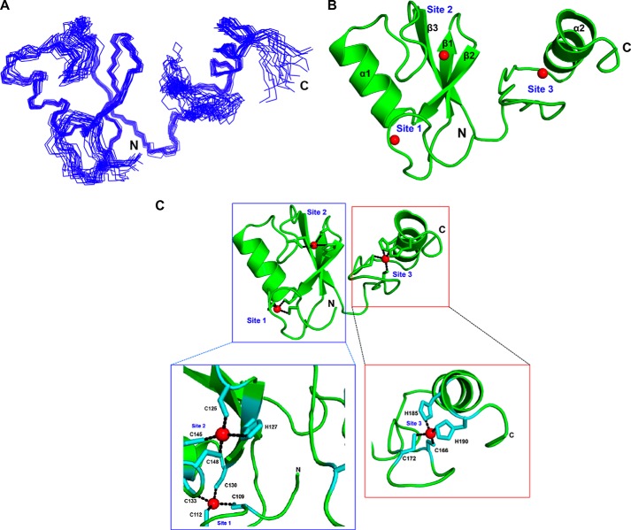
FIGURE 2.
NMR structure of HYBΔC (aa 106–194). A, superposition of 20 energy-minimized conformers representing the NMR structure of HYBΔC. B, ribbon diagram of the lowest-energy conformer representing the three-dimensional NMR structure of HYBΔC. Each monomer contains three zinc-coordination sites: Sites 1, 2, and 3. The zinc ions are shown as red spheres. C, metal coordinations in the NMR structure of HYBΔC. The coordination of zinc ions in the RING domain and the C-terminal domain of HYBΔC are shown.

