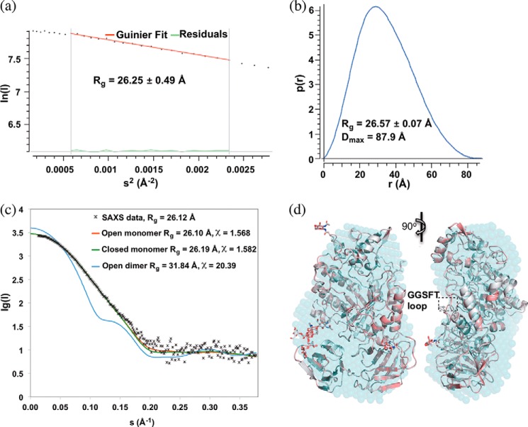
FIGURE 5.
Solution small angle x-ray scattering confirms monomeric structure of FgFCO1. a, p(r) plot. b, Guinier plot. c, fit of experimental SAXS data with theoretical scattering curves for monomer and crystallographic pseudo-dimer calculated with CRYSOL 2.8.2. s = 4πsin(θ)/λ. Both open and closed dimer structures fit poorly (the latter not shown). d, alignment of solution SAXS envelope (cyan dummy atom model) generated by DAMFILT with open (white) and closed (pink) crystal structures of FgFCO1 (secondary structures are shown as schematics and N-glycosylation sugars as sticks). Non-covalently bound ligands and water molecules were removed from the crystal structures during calculation and structure comparison. SAXS data of Endo H-treated FgFCO1 are shown. SAXS data of as-isolated FgFCO1 (not shown) also evidently fit better to monomer structure over dimer structure, showing more heterogeneity than the Endo H-treated sample possibly due to higher and varied degree of glycosylation (Rg values of 27.29 ± 4.87 Å from the Guinier plot and 27.66 Å from the P(r) plot, and Dmax of 95.53 Å; CRYSOL experimental and theoretical Rg values of 27.16 Å and 27.18 Å, and χ of 1.770 for fitting of open monomer structure).

