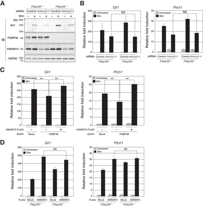
FIGURE 6.
FKBP38 inhibits Shh signaling via suppression of the stimulatory activity of ANKMY2. A, Fkbp38+/+ or Fkbp38−/− MEFs were transfected for 48 h with control or Ankmy2 siRNAs and then incubated in the absence or presence of Shh for 48 h, after which whole cell homogenates were prepared and subjected to immunoblot analysis with anti-Gli1, anti-FKBP38, anti-ANKMY2, and anti-HSP90. B, Fkbp38+/+ or Fkbp38−/− MEFs transfected with siRNAs as in A were incubated in the absence or presence of Shh for 24 h, after which total RNA was extracted from the cells and subjected to RT and real-time PCR analysis of Gli1 (left) and Ptch1 (right) mRNAs. C, MEFs stably expressing EGFP-tagged human FKBP38 and FLAG-tagged mouse ANKMY2, as indicated, were incubated in the absence or presence of Shh for 24 h, after which total RNA was extracted from the cells and subjected to RT and real-time PCR analysis of Gli1 (left) and Ptch1 (right) mRNAs. D, Fkbp38+/+ or Fkbp38−/− MEFs stably expressing FLAG-tagged ANKMY2 were incubated in the absence or presence of Shh for 24 h, after which total RNA was extracted from the cells and subjected to RT and real-time PCR analysis of Gli1 (left) and Ptch1 (right) mRNAs. Data in B–D are means ± S.D. (error bars) from three independent experiments. **, p < 0.01 (Student's t test). NS, not significant.

