
FIGURE 1.
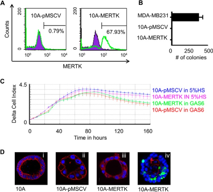
Stable MERTK expression in non-transformed MCF10A cells. A, MCF10A cells were infected with either pMSCV empty vector (left panel) or pMSCV-MERTK (right panel) retroviral particles, and after selection with puromycin, MERTK was detected with anti-MERTK antibodies by flow cytometry. B, MCF10A-pMSCV empty vector control, MCF10A-MERTK, or highly invasive MDA-MB-231 cells were grown in soft agar in the presence of human GAS6, and colonies (where applicable) were stained with crystal violet after 21 days. C, MCF10A-pMSCV and MCF10A-MERTK stable cell lines were starved overnight in medium containing 0.5% HS and full MCF10A supplements. Cells were collected in starvation serum (0.5% HS plus full MCF10A supplements), after which 50 μl of cell suspension containing 2.5 × 10³ cells was added to 100 μl of GAS6 conditioned medium or normal growth medium containing 5% HS and full MCF10A supplements. Real-time proliferation was monitored every 5 h for 160 h by xCELLigence (ACEA Biosciences). D, representative confocal imaging of acini by MCF10A (20× magnification), MCF10A-MERTK (40× magnification), and MCF10A-pMSCV (40× magnification) cells grown in Matrigel for 7 (iv) or 12 (i, ii, and iii) days. Acini were fixed and stained for actin conjugated with Alexa Fluor® 568 phalloidin (red), nuclei with DAPI (blue), and cleaved caspase-3 (green) to identify apoptotic cells (iv).