
Figure 2. 3D reconstruction of cerebellar developmental venous anomaly.
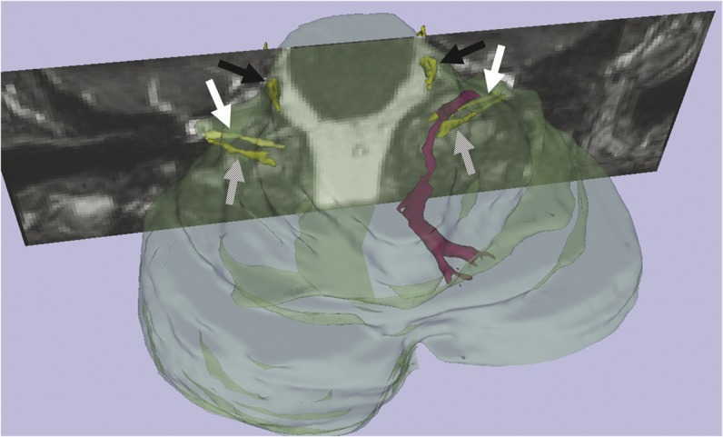
3D reconstructed image shows the developmental venous anomaly and collecting vein (purple) and cranial nerves (yellow): trigeminal, black arrows; facial, white arrows; vestibulocochlear, hashed arrows.