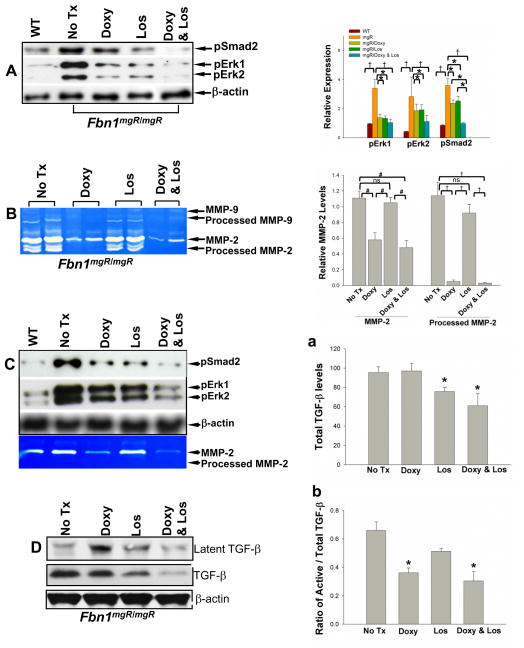
Figure 1.
Phosphorylation of Smad2 and Erk1/2 in the aortic tissues and smooth muscle cells from Fbn1mgR/mgR mice and wild type (WT) littermate controls analyzed by Western blotting (A&C) and MMP-2 and -9 levels analyzed by gelatin zymography (B&C). At the age of 8 weeks, mouse thoracic aortas were harvested from WT control and Fbn1mgR/mgR mice treated without, or with doxycycline (Doxy)(0.5 g/L), or losartan (Los)(0.6 g/L), or doxycycline (0.5 g/L) and losartan (0.6 g/L) added to the drinking water. Aortic proteins were extracted. A) Representative Western blot showing the immunoreactivity to antibodies for phosphorylated smad2 and Erk1/2 (left panel) and relative expression was quantified (right panel) using 5 or more aortic specimens per group; B) A representative gelatin zymogram of MMP-2 and -9 expression using 10 μg of aortic protein per lane with quantification of relative MMP-2 levels in the aortas (n=4) (right panel). C) Aortic smooth muscle cells from wild type and Fbn1mgR/mgR mice were isolated and received no treatment (none), doxycycline (100 μg/ml), losartan (100 μg/ml), or combination of doxycycline (100 μg/ml) and losartan (100 μg/ml). Cellular proteins (25 μg) were analyzed for phosphorylation of Smad2 and Erk1/2 using Western blot (upper panel); MMP-2 expression in cell conditioned media were analyzed by zymography (lower panel). D) TGF-β levels in the aortic tissue of Fbn1mgR/mgR mice with or without treatment were analyzed by Western blotting. Total TGF-β and the ratio active TGF-β to total TGF-β were quantified (n=4) (a&b). Western blots and zymograms are representatives of 3–5 separate experiments. β-actin expression served as internal loading control, * p < 0.05; # p < 0.01; † p < 0.001.

